

Oakstone Clinical Approach to Vascular Ultrasound and RPVI Prep Course 2026
35 $
Format : videos + pdfs, size: GB
Vascular US for Test Prep and More
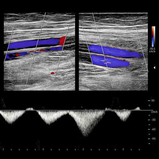
Reading vascular ultrasound is about more than recognizing anatomy. It requires consistent technique, accurate interpretation, and knowing how imaging findings influence patient care. Clinical Approach to Vascular Ultrasound and RPVI Prep Course combines foundation-building insight with real clinical scenarios that walk you through vascular studies from acquisition to interpretation.
Through flexible online video, you’ll review diagnostic criteria, scanning protocols, reporting standards, and common pitfalls while preparing for the RPVI exam. Faculty from Massachusetts General Hospital, Brigham and Women’s Hospital, Stanford, Johns Hopkins, and the University of Colorado cover an array of continuing medical education topics. Highlights include:
RPVI-focused review and exam-tested pathology
Arterial, venous, carotid, cerebrovascular, and abdominal ultrasound
Case discussions spanning routine to complex vascular disease
Quality assurance, reporting, and laboratory standards
Bypass graft, dialysis access, and DVA surveillance
Emerging technologies and new clinical evidence
Date of Original Release: July 31, 2026
Estimated Time to Complete: 13.50 hours
Learning Objectives
Upon completion of this activity, participants will be able to:
Apply standardized, evidence-based vascular ultrasound protocols across arterial, venous, cerebrovascular, and abdominal examinations.
Describe current diagnostic criteria used to interpret vascular ultrasound findings, including velocity measurements and waveform analysis.
Integrate ultrasound findings into clinical decision-making, including determining appropriate intervention, surveillance, or need for additional imaging.
Implement standardized reporting and quality assurance practices consistent with vascular laboratory standards.
Identify and mitigate technical limitations and sources of error to improve study quality and diagnostic reliability.
Intended Audience
Primary care physicians, specialty physicians, physician assistants, and nurse practitioners.
+ Topics:
Basics of Physics in Vascular Imaging – Traci B. Fox, EdD, RT(R), RDMS, RVT, FSVU
Understanding the Ultrasound Machine – Lindsey Ferraro, MS, RDCS, RVT
Peripheral Arterial Studies – Upper – John G. Carson, MD, RPVI, FACS
Peripheral Arterial Studies – Lower – Aarathi Minisandram, MD, MS, MA, RPVI
Abdominal Ultrasound – Scott Manchester, RVT
Panel Discussion – Faculty
Carotid Duplex Ultrasonography – Mingzhu Mary Yan, RVT
Subclavian and Vertebral Ultrasonography – Brian Scholz, RVT, RT(R)
Cerebrovascular Intracranial (Transcranial Doppler) – Sarah LaRose Michaud, RVT
High Yield Pathologies Tested on the Registered Physician in Vascular Interpretation (RPVI) – Abhisekh Mohapatra, MD, MS, MA
Panel Discussion – Faculty
Bypass Graft Surveillance – John G. Carson MD, RPVI, FACS
Aortic Endovascular Stent Graft Surveillance – Kendal Endicott, MD, FACS
Dialysis Access Surveillance and Case Presentations – Elena Foster, MD, RPVI
Tibio – Pedal Access – Lindsey Ferraro, MS, RDCS, RVT
Panel Discussion – Faculty
Venous Ultrasound 1 – Venous Insufficiency, Ultrasound in Office Based Venous Procedures – Julianne Stoughton, MD
Venous Ultrasound 2 – Venous Thrombosis-Obstruction, Pelvic Venous Disorders – Ryan T. Brooks, BS, RVT
Venous Case Presentation – Luis Suarez, MD, FACS, RPVI
Wound Healing in Advanced Limb Salvage – Anahita Dua, MD, MS, MBA, FACS
Carotid Arterial Case Presentations – Drena Root, RVT
Updated Results with the Retrievable System for Drug Delivery – Data from the DEEPER OUS and DEEPER REVEAL Trials – Anahita Dua, MD, MS, MBA, FACS
Peripheral Artery and Bypass Graft Case Presentations – Nikolaos Zacharias MD, MPH, RPVI, FSVS, FACS
Surveillance of Deep Venous Arterialization – (DVA) Grafts – Pre- and Post-Operative Insights and Outcomes – Lindsey Ferraro, MS, RDCS, RVT
Deep Venous Arterialization – Sarah Deery Stasulis, MD, MPH, FACS, RPVI
Panel Discussion – Faculty
Be the first to review “Oakstone Clinical Approach to Vascular Ultrasound and RPVI Prep Course 2026”
You must be logged in to post a review.
Related products


Reviews
There are no reviews yet.