


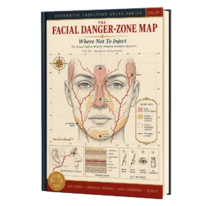
The Facial Danger-Zone Map: Where not to inject, Aesthetic Injection Atlas Series, Vol. 07 (PDF)
6 $
Format Publisher PDF
File size : 61.2 MB
Publisher : Publisher
Published year : 2026
Aesthetic Atlas · The Injectable Series · Vol. 07
The Facial Danger-Zone Map
Where not to inject — mapped, measured, and rehearsed.
Five arteries. Three rescue steps. One map. A 24-page visual field guide to the vessels running under every injection point — the distance to hold, the test to run before the plunger moves, and exactly what to do in the first sixty seconds if the skin blanches.
✓Five arteries mapped anterior and lateral — facial, angular, transverse facial, mental, dorsal nasal
✓The retrograde pathway from the lower face to the ophthalmic circulation, drawn
✓The aspiration test in five steps — fix, pull back, hold 15–30 s, read the hub, decide
✓Recognition and hyaluronidase rescue on one page, ready before you begin
Why this map exists
Distance alone is not safety.
You were taught the numbers — about 1 cm lateral to the facial artery, strictly the middle of the zygomatic arch. Keep them. But a measured gap around a point does not stop material already inside a vessel from travelling the artery’s course.
Warning
Even at the taught distance a bolus can enter the vessel’s course. Aspirate before every injection, without exception.
Key insight
Distance lowers risk; it does not remove it. The vessel’s pathway — not the gap around a point — decides the outcome.
What this guide does
It draws the pathway. Every plate names the vessel, its course, the procedure that endangers it, and the one rule that keeps it safe.
Inside the field guide
Ad-clear at a glance. Clinic-precise on the page.
Large anatomy, short decision notes, and the same page grammar throughout. Six of the twenty-four plates, shown as they print.
01 · Master map
Start with the whole face.
Five arteries on one face, numbered clockwise: facial, angular, dorsal nasal, transverse facial, mental. Then the same danger seen again from the side. Every red line connects — which is why one map governs the whole guide.
Anterior and lateral master plates
Numbered landmark rail, clockwise
Danger corridors in dashed-red hatch
Key insight
Know every vessel before you load a syringe.
02 · Quick reference
Vessel × procedure, on one page.
Six structures, six rules. Facial artery and jawline filler. Angular artery and the tear trough. Transverse facial and the cheekbone. Mental artery and chin boluses. Dorsal nasal and non-surgical rhinoplasty. Parotid gland and the jaw angle.
One row per vessel — where it runs, what endangers it, what keeps it safe
Every cell under fourteen words
The page to photograph before a case
→ Action
Every safe rule reduces to the same three moves: map the vessel, hold your distance, aspirate.
03 · Decision spread
The instrument is a safety decision.
Cannula through the danger corridor for safer transitions and fewer vessels crossed; needle on periosteum where a line must be exact and the product must stay put. The plate presents the trade-off rather than a verdict, because the right answer changes with the corridor.
Mirrored panels, one visual grammar
Strengths and trade-offs side by side
Matched to zone, not to preference
04 · The aspiration protocol
The test, in five steps.
Fix the tip on periosteum. Draw the piston back sharply, several divisions. Hold 15–30 seconds — 30 on the nose. Read the hub. A drop of blood aborts the injection at that point: change the needle, move the point, and never re-inject where the test was positive.
Five panels, left to right, with a progression bar
A companion page on the honest limits of a clean result
Re-test after every needle move
⚠ Warning
“If you see a drop of blood, it means you are in a vessel and you cannot carry out the injection.”
05 · Recognition & rescue
The map continues when something looks wrong.
A sharp pain that does not go away, and a blanched injection site. The plate sets the timeline, then the response: stop injecting and remove the material immediately with high-dose hyaluronidase, capturing 0.5–2 cm around the affected area.
Ischemia timeline with the early signs named
Elective and emergency enzyme use kept visually separate
One-hour monitoring window after enzyme
Key insight
Speed is the treatment. The interval between recognising and flooding the field is the number that matters.
06 · Nerves & glands
Arteries are not the only hazard.
The jaw angle carries two structures at once — the parotid gland and the facial artery. The temple carries a third and fourth: the superficial temporal vein and the temporal branch of the facial nerve, both marked before anything is filled.
Parotid gland and facial artery mapped together at the angle
Temple vein and temporal branch marked before the cannula enters
The temporal crest as a hard ceiling
⚠ Warning
“Dangerous zone. This is parotid salivary gland. It is imperative to perform an aspiration test.”
Be the first to review “The Facial Danger-Zone Map: Where not to inject, Aesthetic Injection Atlas Series, Vol. 07 (PDF)”
You must be logged in to post a review.
Related products





Reviews
There are no reviews yet.