Mayo Point-of-Care Echocardiography: How to Use in Clinical Practice 2020 (Videos + PDFs + Self Assessement)
45 $
Delivery time: Maximum to 1 hours
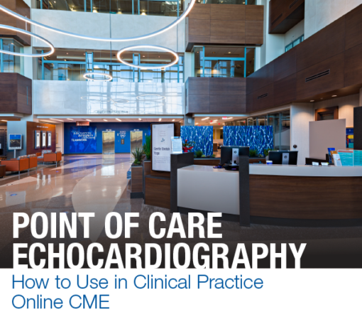
Mayo Point-of-Care Echocardiography: How to Use in Clinical Practice 2020 (Videos + PDFs + Self Assessement)
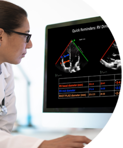
Widespread availability and ease of use of hand-held echocardiography (ECHO) devices is changing clinical practice. This online CME course teaches basic to advanced ECHO to physicians and allied health staff, and includes videos on how to perform ECHO imaging with standard and hand-held systems. The goal of the course to assist learners with prompt bedside diagnoses and ordering appropriate tests. Learners can apply the information to clinical practice, and to expedite patient management and discharge.
Course highlights:
- Didactic lectures by Mayo Clinic echocardiography faculty
- Demonstration of how to perform cardiac and major vascular imaging on the state-of-the-art ultrasound systems by trained cardiac sonographers
- Real-life case examples of myocardial, coronary artery, valvular, pericardial, right heart and aortic pathologies from inpatient and outpatient cardiology consultation practice
- Test your knowledge via multiple-choice questions in individual talks and a separate 47-question quiz.
Target Audience
General cardiologists, hospitalists, intensivists, emergency room physicians, nurse practitioners, physician assistants, and cardiology fellows and residents.
Learning Objectives
Upon completion of this activity, participants should be able to:
- Learn the capabilities, advantages, and pitfalls of currently available hand-held echo devices
- Learn the principles of ultrasound and cardiovascular ultrasound imaging windows
- Understand how to assess significant cardiac, myocardial, valvular, pericardial and vascular pathologies by ultrasound
- Learn how to assimilate information provided by hand-held echo in patient diagnosis and further diagnostic testing needed to assist in patient management
Program :
| Session 1: | ULTRASOUND DEVICES AND IMAGING TECHNIQUES |
| Current Point-of-Care Ultrasound Devices: Imaging Capabilities and Principles of Medical Imaging Tasneem Z. Naqvi, M.D., FRCP(UK), MMM | |
| Cardiac and Vascular Anatomy and Physiology Tasneem Z. Naqvi, M.D. | |
| Ultrasound Physics and Doppler David Pacheco, R.D.C.S. | |
| How to Perform Ultrasound Imaging of the Heart Bobbi J. Heon, R.D.C.S. Deepa R. Mandale, M.B.B.S. Oksana (Semkiv) Ross, R.D.C.S. | |
| How to Perform Imaging of the Cardiac Arterial and Venous Connections Mace H. Ross, R.D.C.S. | |
| Session 2: | CARDIOMYOPATHY AND HEART FAILURE |
| Echocardiography in Coronary Artery Disease Hemalatha (Hema) Narayanasamy, M.B.B.S., M.D. | |
| Coronary Artery Disease – HHE Case Examples Tasneem Z. Naqvi, M.D. | |
| Non-Ischemic CMP Carolyn M. Larsen, M.D. | |
| CMP HHE Case Examples Tasneem Z. Naqvi, M.D. | |
| Session 3: | ASSESSMENT OF RIGHT HEART |
| Echocardiographic Assessment of Right Heart David S. Majdalany, M.D. | |
| Lung Ultrasound Shaun Yang, M.D. | |
| Hand Held Echo Case Examples Tasneem Z. Naqvi, M.D. | |
| Session 4: | VALVULAR HEART DISEASE |
| Mitral Regurgitation Carolyn M. Larsen, M.D. | |
| Aortic Stenosis Tasneem Z. Naqvi, M.D. | |
| Hand Held Echo Case Examples in Valvular Heart Disease Tasneem Z. Naqvi, M.D. | |
| Session 5: | CARDIAC EMERGENCIES |
| Patient with Stroke: Cardiac Thromboembolic Sources Hemalatha (Hema) Narayanasamy, M.B.B.S., M.D. | |
| Pericardial Effusion and Cardiac Tamponade William K. Freeman, M.D. | |
| Aortic Diseases David S. Majdalany, M.D. | |
| Hand Held Echo Case Examples Tasneem Z. Naqvi, M.D. |
Related Products
Video Medical
Emergency Medicine: Evidence-Based Content, Practical Applications 2024 (Videos + Audios)
Video Medical
NYU Langone Health Update on Attention Deficit Hyperactivity Disorder Through the Lifespan 2024
Video Medical
Harvard 9th Annual Board Review and Update in Pulmonary and Critical Care Medicine 2024
Video Medical
Nationwide Children Pediatric Gastroenterology Conference for Primary Care Clinicians 2024
Video Medical
MENA Conference 10th Abu Dhabi International Conference in Dermatology & Aesthetics 2024
Video Medical
Obstetric Anaesthetists Association Joint OAA and UK Maternal Cardiology Society Meeting 2024
Video Medical
Internal Medicine and Urgent Care-Pediatrics: A Review and Update 2025 (Videos + Syllabus)
Video Medical
Obstetric Anaesthetists Association Joint OAA and British Society for Haematology Meeting 2023


